|
|
Medical image quality enhancement |
|
|
Medical image quality enhancement |
|
|
|
|
|
|
|
|
|
|
|
|
|
|
|
|
|
|
|
|
|
|
Previous projects
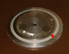
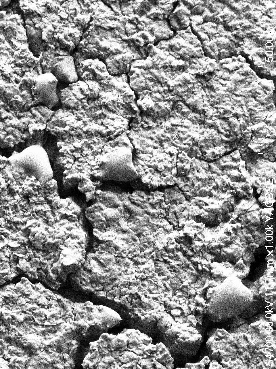
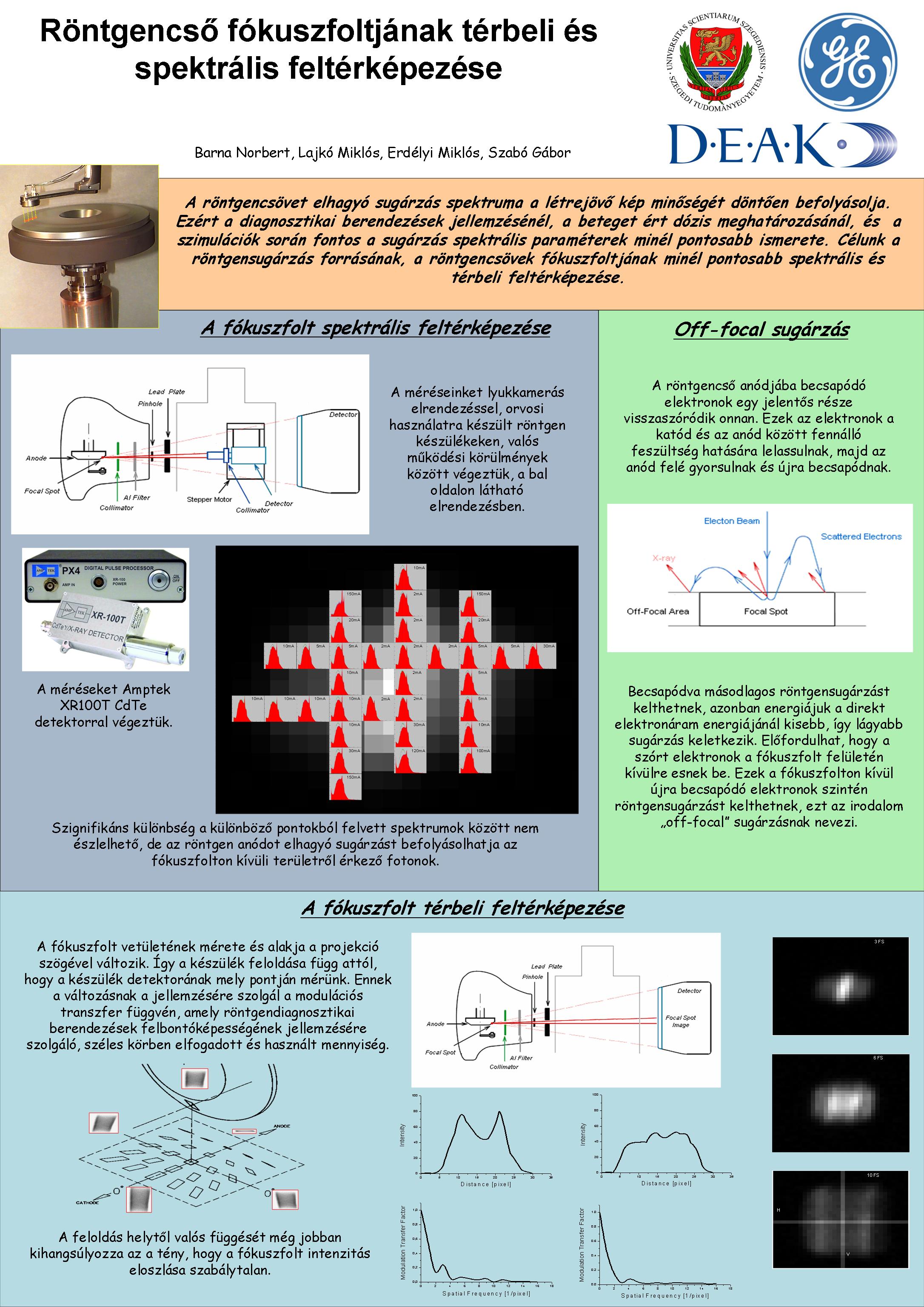
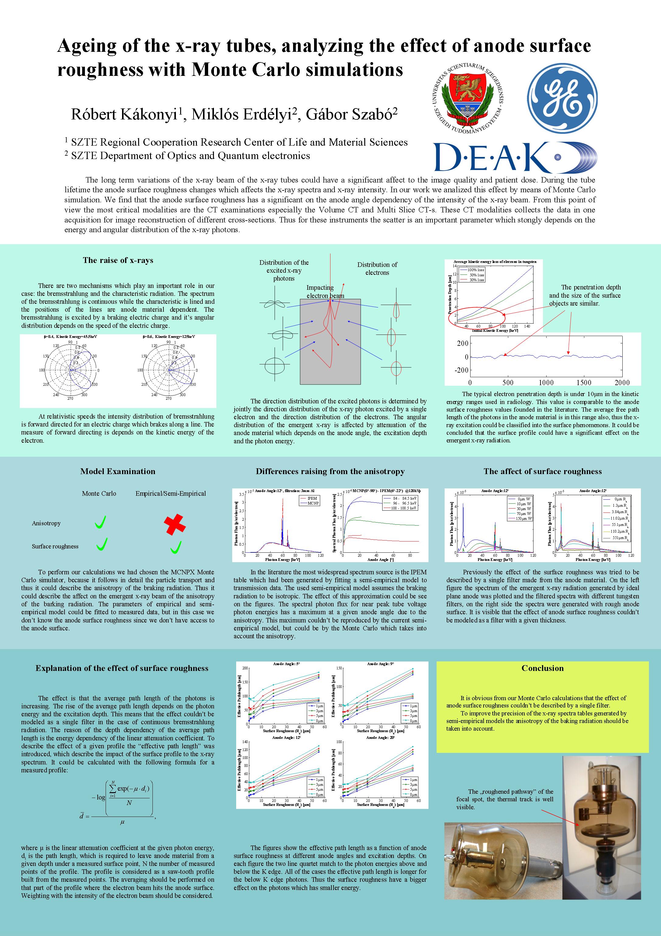
Anode surface roughness measurement:
Heated cathode X-ray tubes are widely used sources in diagnostic medical imaging. In heated cathode tubes X-ray beam is produced by electrons emitted from the cathode. accelerated by the high electric field and collided into the anode surface. As they slow down in the anode material, electrons produce characteristic and bremsstrahlung radiations. However lessthan 1% of the incoming energy electron contribute to the X-ray generation, and the rest is dissipated as heat in the target. The dissipated heat damages the active surface of the anode, generating fissures, tears and cracks limiting the lifetime of the tubes. Besides, the spectrum of the outcoming X-ray also changes (hardening) with the increasing surface roughness.
Measurements with optical microscope, SEM (scanning electron microscope) and profilometer showed that the mud-flatting anode surface consists of island with surface roughness typically smaller than 1 micron. The islands are separated by approximately 8 micron deep tears. Based on the measurement results a simple model was proposed to calculate the modified spectra. Intensity loss of 6% was predicted.

Spectral mapping of the anode and the off-focal area:
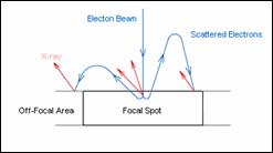
The average penetration of the incident electrons into the anode material is few microns. Significant part of the incident electrons scatter back from the track. These electrons, due to the high voltage between the cathode and the anode, slow down and accelerate toward the anode, and impact to the anode again. After the second impact they can produce secondary X-ray emission. However, their energy is smaller than the energy of the primary electron beam, so softer radiation is produced. Eventually, the backscatter electrons can hit the anode outside the focal spot. These electron also generate X-ray radiation, this radiation is called "off-focal" radiation.
With the knowledge of the spectral distribution of the anode and the off-focal area the system can be better modeled. Spectrum measurements were performed in the area of the anode and the off-focal region using CdTe semiconductor detector, studying the spectral and intensity distribution of the surface of the anode.
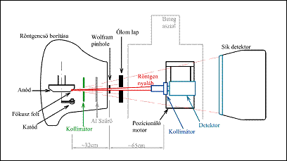
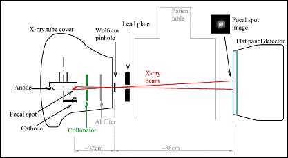
Measurement of the modulation trasfer function of the focal spot:
Depending on the radiological system's composition and on the setup parameters, some blurring appears in the projected images.Part of this blurring is due to the limited resolution of the detecting system and the object moving. Aother part is due to the extended - not punctual - focal spot size and irregular shape. In practice, X-rays do not arise from a point source, but from an extended source with highly irregular shape emission. Chracterization of the diagnostic syystem in this aspect could be useful in many applications.
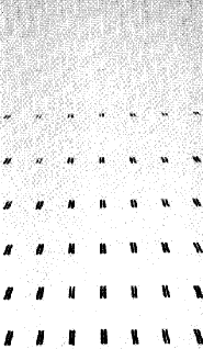
The modulation transfer function (MTF) has become a widely accepted function for the specification of the resolution capabilities of the entire radiological system, groups of components of the system. In radiology, the MTF can be determined with differnet methods, e.g. with star pattern, with slit or pinhole. In our calculations, the MTF curves were determined from point spread functions (PSF) curves measured with pinhole method. The size and shape of the focal spot vary with the angles of projection, hence the radiographic resolution (or the MTF) dependd on the position and the direction of interest in a plane normal to the direction of the beam. Measurements in different postions were performed with the flat panel detector of the vascular system to study the spatial and directinal dependencce of the resolution.
Periodic filtering technique:
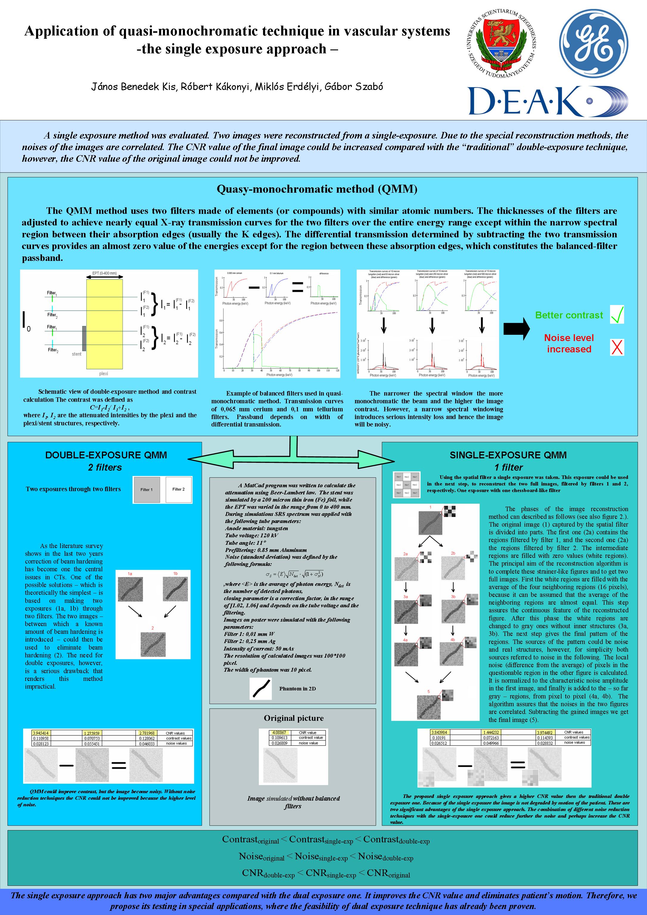
Using two X-ray beams, one can gain information about internal content of the tissues.The dual-energy techniques use two different energy levels to measure, to gain information from the patient. It is usually used to separate soft tissue from the bone. One can get dual-energy information in different ways, using spectral analysization of the detected radiation, or using different tube currents and/or using X-ray filters. With the latter methods, the different energy ranges can be separated very well. Disadvantages of dual exposition (making two different projections) are, the patient moving and the double patient dose.
A new filtering technique had been proposed, which can be used to make the two dual-energy image from one single record. The filter is made of two different materials with a chessboard like shape. The two dual-energy images are obtained by separating the "black" and "white" fileds of the recorded image, and completing images with special algorithms. With this technique artifacts originating from the patient moving can be eliminated, the dose is half than with other dual-energy techniques.
Stent detection by "flashing":
During vascular interventionns, it is extremely important to follow up medical equipments inside the patient's body and to precisely know the postion and the direction of the devices. Such a device is the stent, used to expand the vessel oclusions, which is made of wire and has a web structure.
The idea was that, the moving of the objects helps to the observer to visualizs them. Flashing of an object produces effect like the object woud move. FFT filters were used to flash the stent on the image. The forming frequencies of the stent were determined. The intervals of the stent frequencies are variable video by video, but they were rougly the same. Amplifying the corresponding frequency components in the given frames the visibility of the stents can be enhanced.
Reduction of the effects of scattered radiation on image quality using backscattered photons:
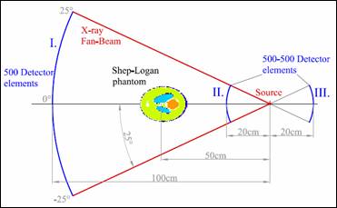
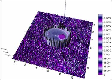
Scattering is a general physical process, the X-ray beam can deflect from its original direction, when travlling through materials. The detection of these scattered photons can cause contrast reduction and corruption of the image quality in CT scanners. The detected scattered radiation consists of the forward scattered radiation, which incides with the same angle to the detector elements, than the original beam. The degree of scattering depends on the metrial of the investigated object, the used geometry, the scanner type, and the used energy range.
Functioning of the CT scanner was modelled with MCNPX ("Monte Carlo") simulator, which is a very powerful and reliable software in the nuclear simulation technique. The images were reconstructed with filtered back projection technique from the the made projections.
Realisation of a new concept was studied, the feasibility of the improvement of the CT images with using backscattered electrons to evaluate the nuber of forward scattered electrons.
Posters:
Articles:
Measurement of X-ray tube anodes' surface profile and its effect on the X-ray spectra
Monte Carlo analysis of energy dependent anisotropy of bremsstrahlung X-ray spectra